

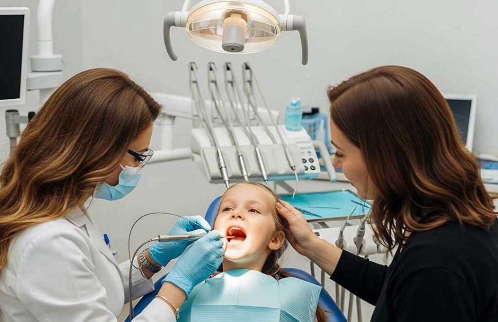
Dental X-rays are diagnostic images that help reveal problems a visual examination cannot always detect. In pediatric dentistry, they may show decay between teeth, developing permanent teeth, changes around tooth roots, jaw growth patterns, or injuries after an accident.
For families in Laguna Hills, understanding why an image is recommended can make the process less stressful. The number and type of X-rays should be based on a child’s age, dental development, symptoms, medical history, and risk of oral disease—not simply on a fixed schedule.
Why might a child need a dental X-ray?
A child may need an X-ray when the information could change diagnosis or treatment. Dental X-rays can help identify:
- Cavities between teeth that cannot be seen directly
- Infection or inflammation near a tooth’s root
- Teeth that are missing, crowded, impacted, or developing unusually
- Damage after a fall, sports injury, or other facial trauma
- The position of permanent teeth beneath primary teeth
- Bone changes, cysts, or other uncommon abnormalities
- The extent of a problem before treatment
Some cavities begin in tight spaces between back teeth. If those surfaces cannot be examined visually or with dental instruments, bitewing X-rays may provide useful information. This is especially relevant once children’s teeth become crowded or their contacts close.
An X-ray is not automatically needed at every visit. A healthy child with low decay risk and visible tooth surfaces may need fewer images than a child with frequent cavities, enamel defects, orthodontic concerns, or a history of extensive dental treatment.
What types of dental X-rays are used for children?
Different images answer different clinical questions.
Bitewing X-rays show the crowns of upper and lower back teeth in one area. They are commonly used to look for decay between teeth and to examine the level of supporting bone.
Periapical X-rays show an entire tooth, including the root and surrounding bone. They may be used when a child has tooth pain, swelling, a history of trauma, or concern about infection.
Occlusal X-rays capture a larger section of the upper or lower jaw. They can help locate an unerupted tooth, investigate unusual development, or evaluate certain injuries.
Panoramic X-rays create a broad image of the jaws and developing teeth. They may be useful for assessing tooth eruption, significant crowding, jaw injury, or overall dental development. Because the image covers a wide area, it does not always show small cavities as clearly as an intraoral image.
Cone-beam computed tomography, or CBCT, produces three-dimensional images. It is not a routine screening tool for most children. It may be considered for selected orthodontic, surgical, trauma, or unusual developmental questions when ordinary dental images cannot provide enough information.
How often should children receive dental X-rays?
There is no single age-based schedule that applies to every child. Imaging frequency depends on clinical findings and the child’s risk profile.
A child with active decay or a higher likelihood of developing cavities may need bitewing images more often than a child with no visible disease and low risk. The timing may also change as primary teeth loosen, permanent teeth erupt, or spaces between teeth close.
A dentist generally reviews:
- The child’s current symptoms and examination findings
- Previous cavities and dental treatments
- Whether tooth surfaces can be seen directly
- Fluoride exposure and oral hygiene habits
- Diet patterns, including frequent sugary or acidic drinks
- Dental development and eruption
- Previous X-rays that may already answer the question
Parents can help by keeping track of recent dental images, especially when a child changes practices or receives care in more than one setting. Sharing earlier records may prevent unnecessary duplication.
Are dental X-rays safe for children?
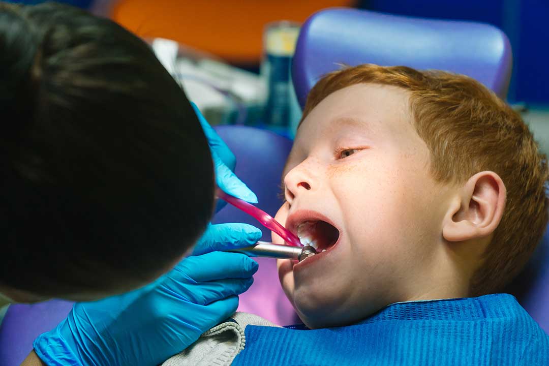
Dental X-rays use ionizing radiation, so they should be taken only when the expected diagnostic benefit justifies the exposure. The amount used for a necessary dental image is low, but children receive special consideration because their bodies are developing and they have more years ahead during which radiation-related effects could potentially appear.
Safety depends on more than the machine itself. Good practice includes using equipment and exposure settings designed for children, limiting the beam to the area being examined, positioning the child carefully, using digital receptors when available, and avoiding repeat images caused by preventable errors.
The guiding principle is often described as ALARA: “as low as reasonably achievable.” In practical terms, this means using the lowest exposure that produces an image clear enough for diagnosis.
Current professional guidance places strong emphasis on accurate positioning, beam restriction, child-sized settings, and avoiding unnecessary examinations. Shielding practices have also been updated in recent years. Families may notice that protective aprons or thyroid collars are used differently depending on the equipment, technique, and current clinical protocol. The central safety goal remains the same: prevent avoidable exposure while obtaining the needed image.
Does a child need to prepare for an X-ray?
Most routine dental X-rays require little or no preparation. A child may be asked to remove eyeglasses, jewelry, hair accessories, or removable dental appliances if they could interfere with the image.
The image receptor or sensor may feel bulky, and some children dislike holding it in position. A brief gagging sensation is possible, particularly for younger children. Calm explanations and simple instructions usually help. Children often cooperate better when they know the process lasts only a few seconds and that staying still prevents the need for a repeat image.
For panoramic imaging, the child generally stands or sits in a fixed position and rests the chin or forehead against supports. The machine moves around the head while the child remains still. Parents should mention developmental conditions, sensory sensitivities, movement difficulties, or previous problems with dental imaging so the process can be adapted appropriately.
Should parents worry about repeated X-rays?
Repeated images deserve a reasonable explanation, but repetition is not automatically unsafe or unnecessary. An image may need to be retaken if the child moved, the teeth were not captured completely, or an important area was obscured.
Parents can ask practical questions:
- What specific concern will this image help evaluate?
- Could an existing X-ray be used instead?
- Is this a routine image or one prompted by a symptom?
- How will the result affect diagnosis or treatment?
- Is a two-dimensional image sufficient, or is three-dimensional imaging being considered?
These questions support informed decision-making without assuming that imaging is either harmless in every situation or dangerous whenever it is recommended.
What should Laguna Hills families remember after an injury?
Children in active households may experience falls, playground accidents, bicycle incidents, or sports-related impacts. After facial trauma, an X-ray may help evaluate a displaced, fractured, intruded, or developing tooth, but not every injury requires the same images.
Urgent dental or medical assessment is especially important when there is uncontrolled bleeding, facial deformity, loss of consciousness, trouble breathing or swallowing, severe swelling, or a permanent tooth that has been knocked out. For less severe injuries, families should still document what happened, save any recovered tooth fragment or tooth as instructed, and provide details about when and how the impact occurred.
Dental X-rays are most useful when they are purposeful, tailored to the child, and interpreted alongside an examination and health history. They are not a substitute for looking at the teeth and mouth, but they can reveal important information hidden beneath the visible surfaces.